|
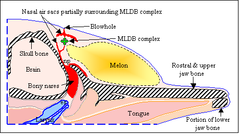
Comments, suggestions, and/or questions are welcomed regarding any portion of the research described here. Email the author at jaroyan@cruzio.com . Simple models of the dolphin echolocation emission system. Click on the link below for an (unfinished) draft of an article on models of dolphin echolocation signal emission. I am hoping that readers will find this draft interesting enough to send their comments and suggestions. This article proposes a simple 3-component acoustical model of the dolphin forehead that appears to explain a great many features of dolphin echolocation signals. The acoustical behavior of each model component is illustrated using simulation movies and examples of model input-output signals are provided. Note: This article contains an introduction to dolphin forehead anatomy and a brief description of dolphin echolocation signals. Readers who are not familiar with dolphin anatomy and/or echolocation signal characteristics may wish to read this introduction before downloading the next article on 2D computer modeling of dolphin echolocation beam formation. I have not yet replaced the figures with thumbnails, so the HTML page with all figures may take several minutes to download (if you are using a modem). HTML version of draft (~1 Mbyte including all figures): Click here to view. The following figure is from the above article draft.
Figure 1. Diagram of selected common dolphin head tissues from the above article. [Figure adapted from Aroyan 1990.] 2D Modeling of acoustic beam formation in the common dolphin, Delphinus delphis. The article below describes how 2D bioacoustic modeling was used to study sonar beam formation by the forehead tissues of the common dolphin. This article describes the methods and summarizes the results of my MS thesis research. Click on the link below for a PDF file. Reference: Abstract: Article PDF file (file size 700 Kbyte): Click here to view. The following figure illustrates a simulation result described in the above article.
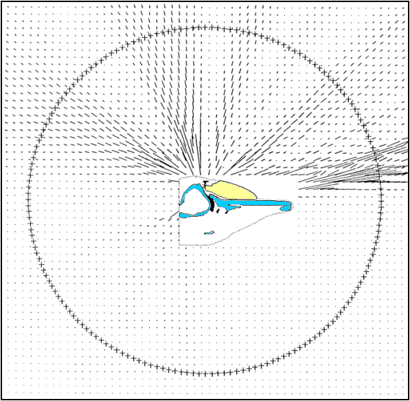
Figure 1. A 2D computer simulation of sound production in the common dolphin. The model of the dolphin's head is indicated by a dotted outline. The yellow region represents the fatty melon tissues, the blue regions indicate skull bones, and the black regions are the nasal air sacs included in the head model. Sonar pulses from a spot (just beneath the uppermost air sac) below the dolphin's blowhole reflect and refract through these structures. The lines around the dolphin's head represent the direction and intensity of sound waves emitted from the model. Most of the acoustic energy is emitted in a forward and slightly upward-directed beam in this 100kHz simulation. The emitted field was collected at the ring of points surrounding the model and compared to the measured beam patterns of live dolphins for several different tissue and source models. Graphic © 1992 Acoustical Society of America. 3D Modeling of biosonar emission in Delphinus delphis. The article below describes the use of 3D acoustic modeling to study sonar signal emission by the forehead tissues of the common dolphin. This research investigated several aspects of the signal emission process in this animal, including the location of the source tissues, focusing by the melon tissues, the focal properties of the skull, and the overall focal characteristics of the complete head of the dolphin. The methods described are more involved than the earlier 2D simulations, and include a novel approach to modeling the acoustic parameters of mammalian tissues based on x-ray CT data. This article appeared as part of a chapter contributed to Vol. 12 of the Springer Handbook of Auditory Research series. It summarizes the biosonar emission methods and results of my PhD research. Chapter Reference: Article Overview: 3D Modeling of hearing in Delphinus delphis. The article below appeared in the December 2001 issue of JASA. This article summarizes the hearing simulation portion my PhD research, describing how 3D bioacoustic modeling was used to investigate pathways of hearing and directional sound reception in the common dolphin. Similar approaches could be used to investigate hearing mechanisms in many other marine mammals. The article notes in conclusion a number of potential refinements and/or extensions of the techniques that may be useful in future applications. Please email me (contact link below) for additional information. I would be delighted to work with sincere collaborators in applying these techniques to other marine mammals. Reference: Abstract: Article draft PDF file (approx. 1.4 Mbyte): Click here to view. The PDF file is actually an author proof of the above article. The color figure resolution has been reduced to minimize file size, but the content is otherwise the same as a reprint. Incidentally, several "puzzling" results of the hearing simulations decribed in the above article have turned out to be supported by more detailed experimental measurements with live dolphins. For example, the simulations indicate that as frequency decreases, the directions of peak hearing sensitivity drop down from the forward horizon to point (at 12.5 kHz) in lateral-ventral directions for each ear. It now appears that this frequency dependence is actually supported in principle by recent jaw-phone sensitivity data for a bottlenose dolphin [see Brill RL, Moore PWB, Helweg DA, Dankiewicz LA (2001) "Investigating the Dolphin's Peripheral Hearing System: Acoustic Sensitivity About the Head and Lower Jaw," SPAWAR Technical Report No. 1865]. A pdf file of Brill et al.'s report is available at the web site: The hearing simulations also showed the same left-right
side asymmetry as Brill et al.'s experimental data. Unfortunately,
Brill et al.'s report was released (to the general public) after
I submitted my final draft, and I did not have an opportunity to
discuss these correlations in the article. 3D Modeling of hearing in Delphinus delphis -- Additional Results Full model (D. delphis) simulated hearing
receptivity plots for frequencies |


